| 3/2013 - 14 |
A Comparison of X-Ray Image Segmentation TechniquesSTOLOJESCU-CRISAN, C. |
| Extra paper information in |
| Click to see author's profile in |
| Download PDF |
Author keywords
image processing, image segmentation, biomedical imaging, digital images, X-rays
References keywords
segmentation(30), image(30), images(11), medical(10), processing(9), analysis(7), techniques(6), automatic(6), active(6), technology(5)
Blue keywords are present in both the references section and the paper title.
About this article
Date of Publication: 2013-08-31
Volume 13, Issue 3, Year 2013, On page(s): 85 - 92
ISSN: 1582-7445, e-ISSN: 1844-7600
Digital Object Identifier: 10.4316/AECE.2013.03014
Web of Science Accession Number: 000326321600014
SCOPUS ID: 84884928131
Abstract
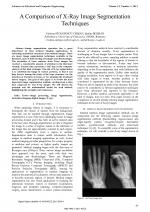
Image segmentation operation has a great importance in most medical imaging applications, by extracting anatomical structures from medical images. There are many image segmentation techniques available in the literature, each of them having advantages and disadvantages. The extraction of bone contours from X-ray images has received a considerable amount of attention in the literature recently, because they represent a vital step in the computer analysis of this kind of images. The aim of X-ray segmentation is to subdivide the image in various portions, so that it can help doctors during the study of the bone structure, for the detection of fractures in bones, or for planning the treatment before surgery. The goal of this paper is to review the most important image segmentation methods starting from a data base composed by real X-ray images. We will discuss the principle and the mathematical model for each method, highlighting the strengths and weaknesses. |
| References | | | Cited By «-- Click to see who has cited this paper |
| [1] V. Zharkova, S. Ipson, J. Aboudarham and B. Bentley, "Survey of image processing techniques", EGSO internal deliverable, Report number EGSO-5-D1_F03-20021029, October, 2002, 35p. [Online] Available: Temporary on-line reference link removed - see the PDF document
[2] D. Feng, "Segmentation of Bone Structures in X-ray Image", PhD thesis, School of Computing National University of Singapore, under guidance of Dr. Leow Wee Kheng (Associate Professor), 2006. [Online] Available: Temporary on-line reference link removed - see the PDF document [3] G. Dougherty. Medical Image Processing Techniques and Applications. Springer, 2011. [CrossRef] [4] J. C. Russ. Image Processing Handbook, the Sixth Edition. CRC Press Taylor & Francis Group, 2011. [5] I. N. Bankman. Handbook of Medical Imaging Processing and Analysis. Academic Press, 2000. [6] J. L. Prince, D. L.Pham, and C. Xu, "A survey of current methods in medical image segmentation ", in Annual Review of Biomedical Engineering, 2:315-338, 2000. [CrossRef] [SCOPUS Times Cited 1967] [7] T. S. Yoo. Insight Into Images Principles and Practice for Segmentation, Registration, and Image Analysis. A K Peters Wellesley, Massachusetts, 2004. [CrossRef] [8] S. V. Kasmir Raja, A. Shaik Abdul Khadir, and S. S. Riaz Ahamed, "Moving toward region-based image segmentation techniques: a study", Journal of Theoretical and Applied Information Technology, 5:81-87, 2009. [9] W. Burgern and M. J. Burge. Principles of Digital Image Processing Fundamental Techniques. Springer, 2009. [CrossRef] [10] J. Bozek, M. Mustra, K. Delac, and M. Grgic, "A survey of image processing algorithms in digital mammography ", Advances in Multimedia Signal Processing and Communications, pp. 631-657, 2009. [CrossRef] [SCOPUS Times Cited 87] [11] P. Meer and D. Comaniciu, "Mean shift: A robust approach toward feature space analysis ", IEEE Trans. Pattern Analysis Machine Intell, 24(5), pp.603-619, 2002. [CrossRef] [SCOPUS Times Cited 10347] [12] B. R. Abidi, J. Liang and M. A. Abidi,"Automatic x-ray image segmentation for threat detection ", Proc. of the Fifth International Conference on Computational Intelligence and Multimedia Applications, 2003. [13] M. Kass, A. Witkin, and D. Terzopoulos, "Snakes: Active Contours ", International Journal of computer Vision, pp.321-331, 1988. [CrossRef] [SCOPUS Times Cited 13710] [14] M. Kulkarni, "X-ray image segmentation using active shape models", Master's thesis, University of Cape Town, 2008. [Online] Available: Temporary on-line reference link removed - see the PDF document [15] H. Mosto, "Fast level set segmentation of biomedical images using graphics processing units", Technical report, Keble College, 2009. [Online] Available: Temporary on-line reference link removed - see the PDF document [16] B. N. Li, C. K. Chui, S. Chang, and S. H. Ong, "Integrating spatial fuzzy clustering with level set methods for automated medical image segmentation", Elsevier - Computers in Biology and Medicine, no.10, pp.1-10, 011. [CrossRef] [SCOPUS Times Cited 414] [17] O. Matei, "Ontology-based knowledge organization for the radiograph images segmentation ", Advances in Electrical and Computer Engineering, 8 (15), pp.56-61, 2008. [CrossRef] [Full Text] [SCOPUS Times Cited 9] [18] M. S. Brown , L. S. Wilson, B. D. Doust, R.W. Gill, and C. Sun , "Knowledge-based method for segmentation and analysis of lung boundaries in chest X-ray images ", Elsevier Computerized Medical Imaging and Graphics, no.2, pp.463-477, 1998. [CrossRef] [SCOPUS Times Cited 94] [19] D. Davis, S. Linying, and B. Sharp, "Neural Networks for X-Ray Image Segmentation ", Proc.of the First International Conference on Enterprise Information System, pp. 264-271, 1999. [20] H. K. Huang, M. F. McNitt-Gray and J. W. Sayre, "Feature selection in the pattern classification problem of digital chest radiograph segmentation ", IEEE Transactions on Medical Imaging, no. 14(3), pp.537-547, 1995. [CrossRef] [SCOPUS Times Cited 119] [21] S. Linying, B. Sharp, and C. C. Chibelushi,"Knowledge-Based Image Understanding: A Rule-Based Production System for X-Ray Segmentation", Proc. of the 4th International Conference on Enterprise Information Systems, vol. 1, pp. 530 - 533, Spain, 2002. [22] I. El-Feghi, "X-ray image segmentation using auto adaptive fuzzy index measure", Proc. of the 47th Midwest Symposium on Circuits and Systems, vol.3, pp. 499-502, 2004. [CrossRef] [23] A. A. Tirodkar, "A Multi-Stage Algorithm for Enhanced XRay Image Segmentation", International Journal of Engineering Science and Technology (IJEST), Vol. 3 No. 9, pp. 7056-7065, 2011. [24] S. K. Mahendran and S. S. Baboo, "Enhanced automatic X-ray bone image segmentation using wavelets and morphological operators", Proc. of the International Conference on Information and Electronics Engineering, 2011. [25] G. K. Manos, A. Y. Cairn, I. W. Rickets and D. Sinclair, "Segmenting radiographs of the hand and wrist", Elsevier Computer Methods and Programs in Biomedicine, vol. 43 (3-4), pp.227-237, 1993. [CrossRef] [SCOPUS Times Cited 35] [26] P. Annangi, S. Thiruvenkadam, A. Raja, H. Xu, X. W. Sun, and L. Mao "A region based active contour method for X-ray lung segmentation using prior shape and low level features", Proc. of the International Symposium on Biomedical Imaging, pp. 892- 895, 2010. [CrossRef] [SCOPUS Times Cited 93] [27] E. H. Said, G. Fahmy, D. Nassar, and H.Ammar, "Dental X-ray Image Segmentation", Proc. of the SPIE, vol. 5404, pp. 409-417, 2004. [CrossRef] [SCOPUS Times Cited 40] [28] Y. Chen, X. Ee, W. K. Leow and T. S. Howe, Automatic extraction of femur contours from hip X-ray images ", Proc. of the First International Workshop on Computer Vision for Biomedical Image Applications, 3765, pp. 200-209, 2005. [CrossRef] [SCOPUS Times Cited 44] [29] C. Ying, "Model-based approach for extracting femur contours in x-ray images", Master's thesis, National University of Singapore, 2005. [30] M. Seise, S. J. McKenna, I. W. Ricketts and C. A. Wigderowitz, "Segmenting tibia and femur from knee X-ray images", Proc. of Medical Image Understanding and Analysis, pp. 103- 106, 2005. [31] H. Chen and A. K. Jain, "Tooth contour extraction for matching dental radiographs" Proc. International Conference on Pattern Recognition, pp. 522-525, 2004. [CrossRef] [SCOPUS Times Cited 54] [32] L. Ballerini, and L.Bocchi, "Bone segmentation using multiple communicating snakes", Proc. of the International Symposium Medical Imaging, 2003. [33] C. J. Taylor, T. F. Cootes and A. Lanitis, "Active shape models: Evaluation of a multi-resolution method for improving image search ", Proc. of the 5th British Machine Vision Conference, pp. 327-336, 1994. [34] G. Zamora-Camarena "Automatic segmentation of vertebrae from digitized X-ray images",PhD thesis, Texas Tech University, 2002. [Online] Available: Temporary on-line reference link removed - see the PDF document [35] G. Behiels, D. Vandermeulen, F. Maes, P. Suetens, and P. Dewaele, "Active shape model-based segmentation of digital X-ray images ", Proc. of the Second International Conference on Medical Image Computing and Computer-Assisted Intervention, pp. 128-137, 1999. [CrossRef] [SCOPUS Times Cited 53] [36] N. Boukala, "Active shape model based segmentation of bone structures in hip radiographs ", Proc. of the International Conference on Industrial Technology, pp. 1682-1687, 2004. [CrossRef] [37] F. Ding, W. K. Leow, and T. S. Howe, "Automatic Segmentation of Femur Bones in Anterior-Posterior Pelvis X-Ray Images", Proc. of the 12th International Conference on Computer Analysis of Images and Patterns, 2007, pp. 205-212. [CrossRef] [SCOPUS Times Cited 25] [38] N. Senthilkumaran and R. Rajesh, "Edge detection techniques for image segmentation - a survey of soft computing approaches ", International Journal of Recent Trends in Engineering, 1, pp. 250-255, 2009. [39] M. A. Ali, L. S. Dooley and G. C. Karmakar, "Object Based Image Segmentation Using Fuzzy Clustering ", Proc. of International Conference on Acoustics, Speech, and Signal Processing, 2006, pp. 105-108. [CrossRef] [40] L. Florea, C. Florea, C. Vertan and A. Sultana, "Automatic Tools for Diagnosis Support of Total Hip Replacement Follow-up ", Advances in Electrical and Computer Engineering, vol.11, no.4, pp.55- 63, 2011. [CrossRef] [Full Text] [SCOPUS Times Cited 6] [41] S. Binitha, S Siva Sathya, "A Survey of Bio inspired Optimization Algorithms", International Journal of Soft Computing and Engineering (IJSCE), ISSN: 2231-2307, vol.2, Issue 2, May 2012. [42] X. Wang, B.S. Wong, C. G. Tui, "X-ray image segmentation based on genetic algorithm and maximum fuzzy entropy", Proc. IEEE Conference on Robotics, Automation and Mechatronics, pp.991-995, 2004. [CrossRef] [43] N. Senthilkumaran, "Genetic Algorithm Approach to Edge Detection for Dental X-ray Image Segmentation", International Journal of Advanced Research in computer Science and Electronics Engeneering, vol.1, no.7, 2012. [44] E. Bonabeau, M. Dorigo and G. Theraulaz. Swarm intelligence. Oxford University Press, 1999. [45] F. Keshtkar, "Segmentation of Dental Radiographs Using a Swarm Intelligence Approach", Proc. of Canadian Conference on Electrical and Computer Engineering, 2006, pp. 328- 331. [CrossRef] [SCOPUS Times Cited 31] [46] T. Sag, M. Cunkas, "Development of Image Segmantation Techniques Using Swarm Intelligence", Proc. of the 1st Taibah University International Conference on Computing and Information Technology, pp.95-100, 2011. [47] A. V. Alvarenga, "Artificial Ant Colony: Features and applications on medical image segmentation", Pan American Health Care Exchanges Conference, pp. 96-101, 2011. [CrossRef] [SCOPUS Times Cited 4] [48] J. Rahebi , H. R. Tajik , "Biomedical Image Edge Detection using an Ant Colony Optimization Based on Artificial Neural Networks", International Journal of Engineering Science and Technology, 2011. [49] S. Gupta, G. S. Sandhu and N. Mohan, "Implementing Color Image Segmentation Using Biogeography Based Optimization", Proc. of the International Conference on Computer and Communication Technologies, pp. 167-170, 2012. Web of Science® Citations for all references: 0 SCOPUS® Citations for all references: 27,132 TCR Web of Science® Average Citations per reference: 0 SCOPUS® Average Citations per reference: 554 ACR TCR = Total Citations for References / ACR = Average Citations per Reference We introduced in 2010 - for the first time in scientific publishing, the term "References Weight", as a quantitative indication of the quality ... Read more Citations for references updated on 2025-07-01 00:54 in 167 seconds. Note1: Web of Science® is a registered trademark of Clarivate Analytics. Note2: SCOPUS® is a registered trademark of Elsevier B.V. Disclaimer: All queries to the respective databases were made by using the DOI record of every reference (where available). Due to technical problems beyond our control, the information is not always accurate. Please use the CrossRef link to visit the respective publisher site. |
Faculty of Electrical Engineering and Computer Science
Stefan cel Mare University of Suceava, Romania
All rights reserved: Advances in Electrical and Computer Engineering is a registered trademark of the Stefan cel Mare University of Suceava. No part of this publication may be reproduced, stored in a retrieval system, photocopied, recorded or archived, without the written permission from the Editor. When authors submit their papers for publication, they agree that the copyright for their article be transferred to the Faculty of Electrical Engineering and Computer Science, Stefan cel Mare University of Suceava, Romania, if and only if the articles are accepted for publication. The copyright covers the exclusive rights to reproduce and distribute the article, including reprints and translations.
Permission for other use: The copyright owner's consent does not extend to copying for general distribution, for promotion, for creating new works, or for resale. Specific written permission must be obtained from the Editor for such copying. Direct linking to files hosted on this website is strictly prohibited.
Disclaimer: Whilst every effort is made by the publishers and editorial board to see that no inaccurate or misleading data, opinions or statements appear in this journal, they wish to make it clear that all information and opinions formulated in the articles, as well as linguistic accuracy, are the sole responsibility of the author.


